Atlas of Ultrasound in Obstetrics and Gynecology 3ed 2018

Atlas-of-ultrasound-in-obstetrics-and-gynecology-3ed-2018

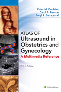
Packed with more sonographic images than ever before, the third edition of the Atlas of Ultrasound in Obstetrics and Gynecology helps you better understand and interpret sonographic imagery, and improve diagnostic accuracy of ultrasound images. This comprehensive visual tutorial is split into two distinct sections: obstetrical ultrasound and gynecological ultrasound. Each section covers normal and abnormal anatomy, pathology, and interventional procedures.

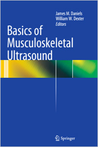
Basics of Musculoskeletal Ultrasound 2ed 2021

Basics-of-musculoskeletal-ultrasound-1ed-2013

Basics of Musculoskeletal Ultrasound, 2e is a must-have reference for residents, fellowship directors, fellows and primary care physicians as well as athletic trainers, physician assistants, physical therapists and ultrasound technicians. It is also an excellent resource for participants of the AMSSM MSK ultrasound courses.

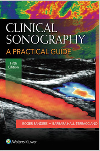
Clinical Sonography: A Practical Guide 5ed 2015

Clinical-sonography-5ed-2015

Uniquely organized by symptom rather than organ or pathology, Roger Sanders’s Clinical Sonography, 5e, not only ensures mastery of the content and competencies required for diagnostic sonography, it teaches students to think critically and “sonographically” as they prepare for certification exams and clinical practice.

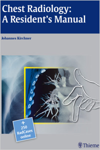
Chest Radiology: A Residents Manual 1ed 2011

Chest-radiology-a-residents-manual-1ed-2011

Chest Radiology: A Resident’s Manual is a comprehensive introduction to reading and analyzing radiologic cardiopulmonary images. Readers are guided through systemic image analysis and can further enhance their learning experience with training cases found at the end of each chapter.

Basics of Musculoskeletal Ultrasound 1ed 2013

Basics-of-musculoskeletal-ultrasound-1ed-2013

Basics of Musculoskeletal Ultrasound speaks to this audience by combining practical clinical aspects, from selecting the proper ultrasound machine to “knobology” and tissue scanning, to specific information organized by anatomical structure. By following this very easy step-by-step guide, the clinician can easily reference the patient’s position, transducer position and the structure to be scanned, and all instructions correspond with recently published ultrasound guidelines.​

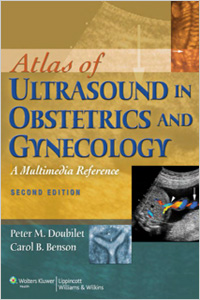
Atlas of Ultrasound in Obstetrics and Gynecology 2ed 2011

Atlas-of-ultrasound-in-obstetrics-and-gynecology-2ed-2011

Atlas of Ultrasound in Obstetrics and Gynecology (second edition) is ideal as a visually-oriented reference and comprehensive tutorial for expanding your knowledge of ultrasound. Developed from one of the best and broadest sonographic collections in the field, this powerful atlas provides images and authoritative commentary covering all aspects of obstetric and gynecologic ultrasound.

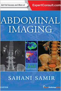
Abdominal Imaging 2ed 2017

Abdominal-imaging-2ed-2017

Richly illustrated and comprehensive in scope, Abdominal Imaging, 2nd Edition, by Drs. Dushyant V. Sahani and Anthony E. Samir, is your up-to-date, one-volume source for evaluating the full range of diagnostic, therapeutic, and interventional challenges in this fast-changing field. Part of the Expert Radiology series, this highly regarded reference covers all modalities and organ systems in a concise, newly streamlined format for quicker access to common and uncommon findings.

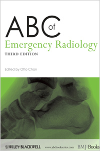
ABC of Emergency Radiology 3ed 2013

Abc-of-emergency-radiology-3ed-2013

The ABC of Emergency Radiology is a simple and logical step-by-step guide on how to interpret radiographs, US and CT. It incorporates all the latest technological advances, including replacing plain radiographs with digital radiographs, changes in imaging protocols and the role of portable US and multidetector CT.

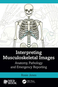
Interpreting Musculoskeletal Images: Anatomy, Pathology and Emergency Reporting 1Ed 2023

Interpreting-Musculoskeletal-Images-1Ed-2023

Sách hướng dẫn giải thích và báo cáo hình ảnh cơ xương khớp, bao gồm các bệnh lý chấn thương thông thường, cơ chế chấn thương, hệ thống phân loại, bệnh lý khớp. Được minh họa đẹp mắt với sơ đồ, hình vẽ và bổ sung bằng hình chụp X quang, nội dung đã được phát triển để nâng cao việc học và hiểu biết về cả X quang và giải phẫu cũng như mối quan hệ giữa chúng.