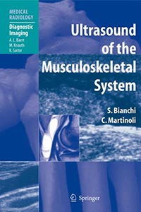
Ultrasound of the Musculoskeletal System 1ed 2007

Ultrasound of the Musculoskeletal System 2007

A comprehensive reference and practical guide on the technology and application of ultrasound to the musculoskeletal system.
It is organized into two main sections. The first is devoted to general aspects, while the second provides a systematic overview of the applications of musculoskeletal ultrasound in different areas of the body.

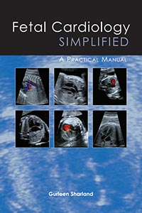
Fetal Cardiology Simplified: A Practical Manual 1ed 2013

Fetal Cardiology Simplified A Practical Manual

Fetal cardiology has developed dramatically into a subspecialty in the past 25 years. The majority of people examining the fetal heart are not ‘experts’ in fetal cardiology and therefore find interpreting images, particularly in case of abnormality, rather difficult. This book is designed as a practical guide, to be kept near the ultrasound machine, for all those performing fetal heart scans, but without the expertise of a fetal cardiologist. The aim is to allow the user to recognize the common forms of fetal cardiac abnormality and to appreciate the associated lesions and outcome.

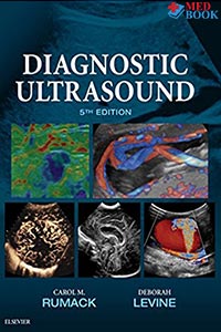
Diagnostic Ultrasound 5ed 2018 Carol Rumack

Diagnostic Ultrasound 5ed Carol Rumack

Now fully updated with more than 2,000 new images, 200 new videos, and new content throughout, Diagnostic Ultrasound, 5th edition, by Drs. Carol M. Rumack and Deborah Levine, remains the most comprehensive and authoritative ultrasound resource available. Spanning a wide range of medical specialties and practice settings, it provides complete, detailed information on the latest techniques for ultrasound imaging of the whole body; image-guided procedures; fetal, obstetric, and pediatric imaging; and much more. Up-to-date guidance from experts in the field keep you abreast of expanding applications of this versatile imaging modality and help you understand the “how” and “why” of ultrasound use and interpretation.

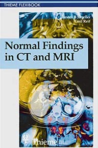
Normal Findings in CT and MRI 1ed 2000

Normal Findings in CT and MRI

The key for any beginning radiologist who wishes to recognize pathological findings is to first acquire an ability to distinguish them from normal ones. This outstanding guide gives beginning radiologists the tools they need to systematically approach and recognize normal MR and CT images.

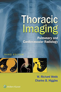
Thoracic Imaging: Pulmonary and Cardiovascular Radiology 3ed 2016

Written by two of the world’s most respected specialists in the field, Thoracic Imaging: Pulmonary and Cardiovascular Radiology, Third Edition brings you completely up to date with all you need to know for optimal imaging of the heart and lungs. This comprehensive title provides practical, authoritative guidance for the radiologic assessment and diagnosis of both congenital and acquired cardiovascular and pulmonary diseases. A must-have reference for more than ten years, Thoracic Imaging is your one-stop source for current, accurate coverage of pulmonary infections, diffuse lung diseases, mediastinal masses, coronary artery CT, myocardial disease, pericardial disease, CT of ischemic heart disease, and much more.

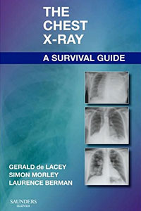
The Chest X-Ray: A Survival Guide 2008

Featuring a practical, clinical approach – and written in a quick-access style – this portable, economical reference helps you build a strong foundation in chest x-ray interpretation. Three radiologists with years of clinical and teaching experience present fundamental principles and key anatomical concepts…walk you through examples of classic chest x-ray features that provide subtle evidence of abnormality…and explore a variety of problems and dilemmas common to everyday clinical practice. High-quality drawings and digital chest x-rays – combined with secrets from the radiologists’ toolbox, helpful differential diagnoses, handy checklists, and key references – deliver all the assistance you need to enhance your interpretation skills.

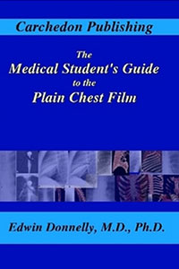
The Medical Student’s Guide to the Plain Chest Film 1ed 2006

This book is designed to provide the medical student a simple yet systemized approach to interpreting the plain chest radiograph. The book teaches the normal anatomy as well as a systematic approach that can (and should) be used to evaluate every chest film. Full-color volume-rendered computed tomography (CT) images are used to help the student understand the anatomy.

Imaging of Cerebrovascular Disease: A Practical Guide 1Ed 2016

Imaging plays an integral role in the diagnosis and intervention of debilitating, often fatal vascular diseases of the brain, such as ischemic and hemorrhagic stroke, aneurysms, and arteriovenous malformations (AVMs). Written by a world renowned neuroradiologist and pioneer in the early adoption of magnetic resonance (MR) technology, Imaging of Cerebrovascular Disease is a concise yet remarkably thorough textbook that advances the reader’s expertise on this subject.